Software for Digital microscopy

Learn More About Us
our-business

Our business

For pre- and clinical research and diagnostic purposes, it’s important that medical images are not just images, diagnosis reports are not just unstructured information, molecular data are not just separated from results of image analysis. Researchers would be supported best, if suspicious or malicious structures of tissue samples are identified at the early stages. In that case the researchers and doctors be able to apply their expert knowledge more efficiently and to focus on the illness itself.
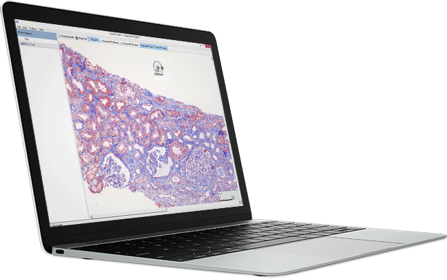
This is exactly, what the HS Analysis GmbH from Karlsruhe (GER) does science 2015, when it comes to automated image analysis, structure reports and bring them with knowledge from molecular data. The integration of these approaches in the hardware and its infrastructure is one of the main skills of HS Analysis.
The company is working worldwide closely with different partners together, when digitization, digitalization and automation of medical data is going to be a part of daily routine in diagnosis and research with AI techniques.
With the help of modern AI, Deep Learning and Active Learning supported methods HS Analysis, believes to seriously inspire quantification and prediction in diagnosis, CDx and research areas such as the evaluation or early detection of e.g. cancer.

technology

IoT Powered Microscopy at Inexpensive Pricing

The most up-to-date artificial intelligence is one key technology of automatic interpretation of tissue samples in the software of HS Analysis. Technically speaking, virtual slides are analyzed with deep learning. Virtual slides are digital images, that are produced by microscope roboters called slide scanner. In this way you quickly get quantitative results about effects of drug candidates on tissue.

Specialists of HS Analysis use the appropriate technology depending on requirements of customers.
Two examples of most used technologies are listed below.

Digital microscopy is a promising method for quantitative automatization in pharmaceutical research. The key technology was the introduction of Whole-Slide Imaging (WSI) or well plates with digital slide Scanners or HCS. They allow virtualization of the whole slide or well plate with large tissue sections or cell cultures in high microscopic resolution. Combination of WSI or well plates with automatic detection and quantification software enables numerous new possibilities and methods to search for drugs against diseases. HS Analysis develops automatic WSI and well plates quantification solutions for efficient workflows in pharmaceutical research. Our solutions are compatible with all slide scanner or HCS file formats.
Recent evolution in artificial neural networks allows reliable recognition of structures with complex morphology traits, similar like the human brain does. HS Analysis specialized in neural network applications and Deep Learning to detect your Region of Interest in automatic manner. Avoid manual annotations for ROI definition to achieve high throughput with short analysis times.

details

Details

HSA cell-tracking & analysis in real-time

HSA deep learning


Tracking of cellular morphology changes and behavior in concert with surrounding cells. Cells with increasing area are marked blue, decreasing area red. Length of individual cell borders, circumference, area and percentile change, compared to previous frame (Δ area, Δ circum.), are tracked in real time.

HSA Deep Learning:
  • + Cell border gaps in the original image are closed intelligently
  • + Automatic tracking of length of individual cell borders, cellular morphology changes and behavior in concert with surrounding cells
  • + Visual representation of selected morphology changes (Area, circularity, neighbor cell behavior etc.)
  • + Simple export of all statistical data into an Excel-file

Standard cell-tracking

Standard U-Net Deep Learning


Simple intensity dependent annotation of cell borders vs original image


Standard U-Net deep learning:
  • – Gaps in the original image are not closed, therefore subsequent statistical analysis is impaired
  • – Tracking of cellular parameters difficult or only possible by additional programs
  • – No visual representation of selected changes in cellular morphology


Counting and segmentation of
cells in chamber slides

Detection and segmentation of nuclei and cytoplasm in cultured cells in chamber slides.


Segmentation of Tumor
and Stroma cells

Segmentation of nuclei and cytoplasm in tumour cells and cytoplasmic regions in stroma cells. If nuclear staining is not present or unspecific, nuclei can be detected by nuclear “holes” in cytoplasmic staining.


Kidney Tool

Segmentation of glomeruli and tubuli by using a Deep Learning model for brightfield data.


Kidney Tool

Segmentation of glomeruli and their compartments by using a tensorflow model for brightfield data.


Cell Counting

Segmentation and counting of cells.  


Endoplasmic Reticulum

The endoplasmic reticulum (ER) is a dynamic structure consisting of branched domains and tubular sections. Automatic segmentation opens up new possibilities for quantitative analysis and efficient representation of this membrane structure at the ultra-structural level.


Scratch Assay

Analysis of samples with scratches and the change of cell area over time.


Internalization

Analysis of insulin receptors inside and outside of cytoplasm and the change over time.


Blood vessel analysis

Blood vessels can be marked with immunohistochemistry. This allows blood vessel counting and analysis of blood vessel distribution.


Real time detection on microscopy devices

You look through the microscope and see in real time markings of the objects relevant to you. Subsequently, an automatic quantification of the detected objects takes place.


Proliferation

Segmentation of cells over time to measure proliferation. The analysis is done on Label Free Data.


Colorectal cancer

Segmentation of brightfield data with colorectal cancer using a deep learning model.


Lung analysis

Automated classification of tumors in non-small cell lung cancer (NSCLC) and quantification of tumor/stroma.


Intracellular Compartmentally in EM

The ultrastructural analysis of intracellular compartments using electron microscopy provides new insights into cellular Fine structures and subcellular organelle interactions.


Annotation Software to set region of interests (ROI)

Manual drawing of regions of interest (ROI) as well as automatic ROI detection. Image analysis can be restricted to ROIs or different analysis methods can be allocated to different ROI types within one image.


Screening with oncology markers

Tissue evaluation with cancer specific markers, for example PD-L1, Ki67, p21, p53, Stathmin, bTubulin, etc.


TMA Screening & Analysis

Analysis Software for Tissue MicroArrays (TMA) with variable number of cores. Semi automatic naming of TMA cores. Export of TMA core results together with cores names into excel files.

congress

Events

Pictures

Fotos mainly by: Michael M. Roth


Job Offers (Students & Professionals)

You will find us here:

Netzwerk Deutschlandstipendium Karlsruhe Logo
27th January 2020
3 founders talk about their experience of starting a business and report from different phases of the foundation
Great Hall, Gastdozentenhaus,
Engesserstraße 3, 76131 Karlsruhe

More Information

Industrie & Innovationstag Logo
28th February 2020 from 4:00 pm to 6:00 pm
3rd Industry & Innovation Day.
Internationales Wissenschaftsforum Heidelberg (IWH),
Hauptstraße 242, Heidelberg

More Information

Scion Logo
23-25th October 2020
Scion : Vertrauen in KI
Technologiefabrik – Haid-und-Neu-Straße 7, Karlsruhe

More Information

past events

Scion Logo
30th November 2019
Scion: Vertrauen in KI
Technologiefabrik – Haid-und-Neu-Straße 7, Karlsruhe

More Information

HS Pforzheim Logo
November 14, 2019
You are the future of tomorrow!
On the way to practical experience with Deep Learning in Life Science.
University of Applied Sciences Pforzheim,
Medical Imaging

Weitere Informationen

BioRN Logo
November 11, 2019
The BioRN Annual Conference "Artificial Intelligence meets Health" will take place on 11.11.2019 in the DKFZ.

More Information

Nachfolgeforum Logo
October 15, 2019
At the Successor Forum 2019 we talk about Artificial Intelligence and implications for successor design.
Europa Park, Rust

Weitere Informationen

AHK
October 11, 2019 at 11:00
Lecture: How autonomous are the autonomous AI systems?
Delegation Deutsch-Baltische Handelskammer (AHK)
Technologiefabrik Karlsruhe

Weitere Informationen

Rotary Club Pforzheim
26th of September 2019
Lecture: Trust in AI - myth or reality?
Business Forum at the Rotary Club in Pforzheim

Weitere Informationen

September 26, 2019
Creating confidence in the AI and securing profitability for SMEs.
Business Forum Ettlingen

Weitere Informationen

Zukunftstüftler Logo
September 18, 2019
127/5000 Future inventors - the conference for the minds of digital transformation
in the Technikfabrik Karlsruhe

Weitere Informationen

W3 Fair
19th of September 2019
Lecture: Trust in AI - myth or reality?
Dornbirn (Vorarlberg)

Weitere Informationen

BioLAGO Logo
July 7, 2019
Seven creative designers from Karlsruhe have seven minutes to present themselves and their company.

Event on Facebook
More Information

BioLAGO Logo
June 27, 2019
BioLAGO „Drugs, Diagnostics, Devices & Data – Matchmaking with Ireland”
Vortragsthema: „Künstliche Intelligenz: Deep Learning in der Medizin, Mythos oder Realität?“

More Information

20 May 2019
2nd European Pathology Congress in Berlin
Vortrag zum Thema Opportunities and risks of AI integration in daily routine in pathology today

More Information

Etengo-Symposium
16 May 2020
Etengo-Symposium 2019
Workshop zur Anwendung von Deep Learning in Bildanalyse
WIRSOL Rhein-Neckar-Arena in Hoffenheim

More Information

DBT
April 9, 2019, 10-17 clock
Deutschen Biotechnologietage (DBT) Symposium
Vortrag zum Thema Bio-Daten und Deep Learning
In Würzburg

More Information

Scion
October 26-28, 2018
2: Scion : Vertrauen in KI
Technologiefabrik – Haid-und-Neu-Straße 7, Karlsruhe

More Information

House of Pharma
September 11, 2018
Auf der Spitze des Hype Cycles: Wie Deep Learning die Entwicklung neuer Wirkstoffe und Therapien mit signifikanter Zeit- und Kostenersparnis ermöglichen wird.
Frankfurt am Main

More Information

IHK
5 July 2018, 2-6 pm
Erster Tag der Künstlichen Intelligenz
IHK Karlsruhe

More Information

Scion
23 + 24 June 2018
1: Scion : Vertrauen in KI
Technologiefabrik – Haid-und-Neu-Straße 7, Karlsruhe

More Information

Steinbeis
Friday, June 15, 2018
Maschinelles Lernen: Produktionsanwendungen in der Robotik
ZKM – Zentrum für Kunst und Medien, Karlsruhe

More Information

CEBIT
June 14, 2018
CEBIT
Hannover

More Information

Michelin
June 12, 2018 16:00 - 20:00
Qualitätssicherung mit VR und KI
Michelin Reifenwerke Karlsruhe

More Information

Our Blog:

front-page

Drop us a line!