Introduction:
Ocular tumors are tumors inside the eye. They are collections of cells that grow and multiply abnormally and form masses. They can be benign or malignant. Specialists at HS Analysis truly believe in better healthcare and treatment of diseases in ophthalmology by using AI algorithms as guided tool in hospitals and research centers. Our software and Hardware system continues to support experts in the Medical field by analyzing medical data in ophthalmology by distinguishing tumor from non-tumor.
Modalities used for diagnosis
The world of intraocular imaging is exploding with new technology. For years, fundus photography, fluorescein angiography, and ocular ultrasonography prevailed, but now there are even more choices, including microimaging modalities such as optical coherence tomography (OCT), OCT angiography (OCTA), fundus autofluorescence (FAF), and indocyanine green angiography (ICGA). There are also the macroimaging technologies computed tomography (CT) and magnetic resonance imaging (MRI). Yet to be explored fully for use with intraocular tumors are multispectral imaging, dark adaptation, and adaptive optics. With all of these refined tools, how is a clinician to choose the appropriate one?
the most effective tool for assessing intraocular tumors is indirect ophthalmoscopy in the hands of an experienced ocular oncologist. Numerous factors go into the equation of tumor diagnosis, such as lesion configuration, surface contour, associated tumor seeding, presence and extent of subretinal fluid, shades of tumor coloration, intrinsic vascularity, and others that hint at the diagnosis and direct our thoughts on management. For example, an orange-yellow mass deep to the retinal pigment epithelium (RPE) or in the choroid with surrounding hemorrhage and/or exudation would be suspicious for peripheral exudative hemorrhagic chorioretinopathy (versus choroidal metastasis from renal cell carcinoma or cutaneous melanoma). The combination of features leads to pattern recognition.
Detection and analysis
HS Analysis’s software is a useful tool to aid in the early detection and analysis of eyelid tumors. With its assistance, primary care providers and ophthalmologists can potentially improve their ability to differentiate between tumors and non-tumor conditions, ultimately leading to earlier diagnoses and more effective treatment options.
The software use various techniques, such as image recognition algorithms and machine learning models, to identify and classify tumors based on characteristics such as size, shape, and location. It also help to compare images of a patient’s eyelids over time to detect any changes that may indicate tumor growth or progression.
Overall, the development of HS Analysis’s software represents a promising step towards improving the early detection and analysis of eyelid tumors, which ultimately lead to better patient outcomes and a reduction in morbidity and mortality.
Importance of Ophthalmic tumor in HSA software:
•The technician can Classify few tumor cells so they can be used as ground truth data (GTD) for the training of a Deep Learning model.
• By using AI algorithms incorporated into the program, that quickly locates and categorizes tumors using Deep Learning models.
• It makes early detection of tumors fast and more accurate.
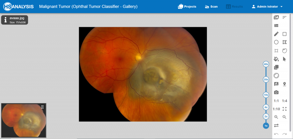
The way Ophthalmic tumor model functions:
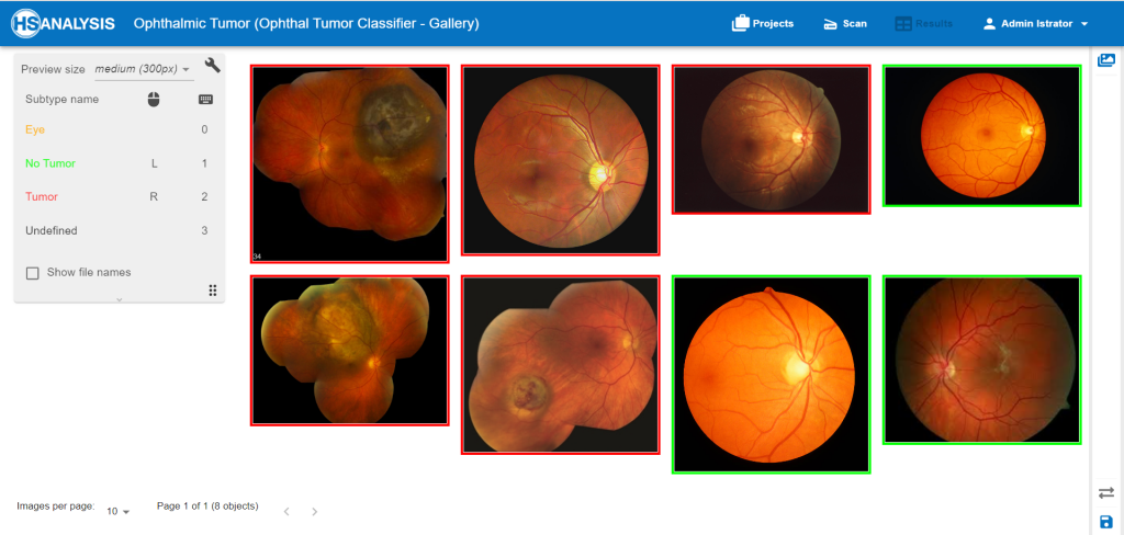
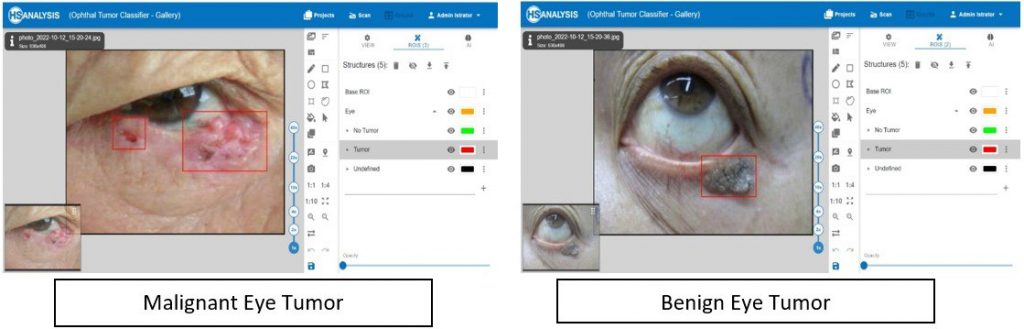
• we simply categorize which images include tumors and which do not, as seen in the image above.
• Then the program develops a Deep Learning model which can automatically recognize tumors in other images.
• The non-tumor and tumor eyes are automatically distinguished using our DL model
State of the art of Classification architectures:
HyperTumorEyenet(Type1) was implemented that is based on EfficientNet, which is a convolutional neural network (CNN) architecture, that uniformly scales the depth, width, and resolution using a fixed set of scaling coefficients, rather than scaling them arbitrarily as is commonly done.HyperTumorEyenet(Type1) was implemented by using integrated feature built-in within the HSA KIT software.
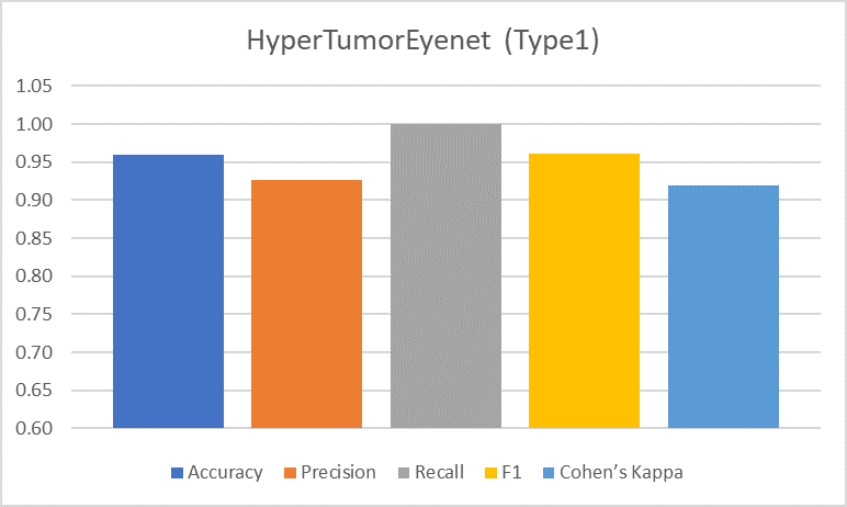
The results show that the model achieved an accuracy of 96%Additionally, it shows that the precision of the model was 92.59%, . Furthermore, the model achieved recall of 100% , F1 score also achieved 96.15%, and Finally, the Cohen Kappa score reached 92%.
xAI in HSA KIT software:
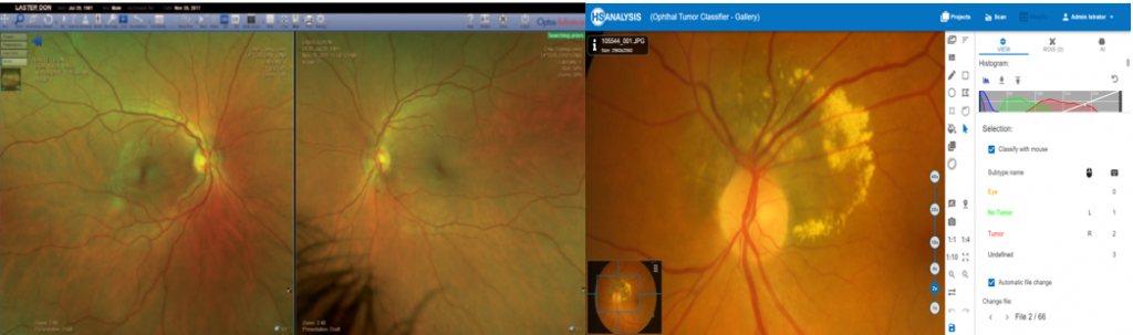
HSA KIT software technique for xAI was used, which is used to locate class specific image region.
The steps taken to implement xAI with HSA KIT Software technique include: loading an image, pre-processing the image, loading the pre-trained model, passing the processed image through the model to generate predictions, defining the target class, computing gradients, applying global average pooling, generating the heatmap, and finally, overlaying the heatmap on the original image to visualize the areas contributing to the target class prediction.
AS a HSA KIT customer you can understand the decision of already trained deep learning model as well as understand which direction should be optimized to get best quality deep learning model during the training process.in this iterative process of training deep learning model you can see exactly which features should be activated or how much more data of specific class should be used to get best quality results, as Figure below shows tumor images as identified by the model; warm colors (yellow, red) signify regions within the tumor tissue sample that exhibit a higher concentration/intensity of tissue activity as detected by the model.

The results of xAI showed that software method accurately identifies regions of high tissue activity, which are represented with warm colors, The results indicate that the performance of HyperTumorEyenet(Type1) yielded accurate and precise results, making it an excellent option for medical diagnosis.
HS Analysis interoperable with Hardware/Software devices
HSA KIT matches perfectly with the following devices to work on Ophthalmic solutions
FF 450plus
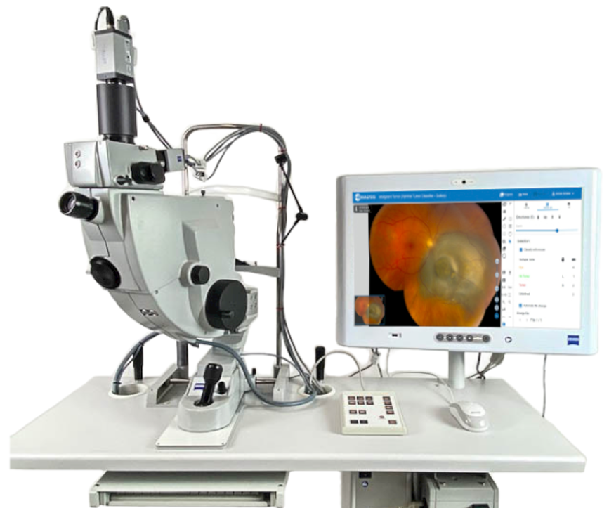
The medical experts working with this device on retinal diagnostic procedures which produce a unique level of diagnostic accuracy, and deliver images with the clearest details, thus providing a dependable basis for first-class treatment outcomes.
Combined with HSA’s deep learning by using AI algorithms incorporated into the program software kit it allows the medical professionals to work more efficiently on the diagnostic procedure and reach end results faster and more accurately.
CLARUS 700
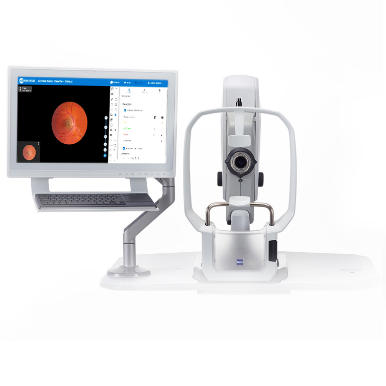
This device from ZEISS was developed as a comprehensive ultra wide-angle fundus camera for ophthalmologists. This enables ultra-wide-angle images to be recorded in true color and with first-class image quality. It offers the full range of imaging modalities, including fluorescence angiography.
Output from this device can be analyzed using HSA kit to annotate and highlight tumor cells and abnormal tissue faster using deep learning algorithms.
Optos Panoramic Ophtaloscope P200Tx
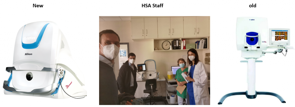
This confocal scanning laser ophthalmoscope is a widefield digital imaging device that can capture images of the retina from the central pole to the far periphery. The retinal images are captured automatically and in a patient-friendly manner, with no scleral depression or contact with the cornea.
The images captured by these devices (New,Old) will be scanned and analyzed using the HSA kit to generate a highlighted and annotated output image of tumor cells.
Heidelberg Engineering HRA + OCT Spectralis
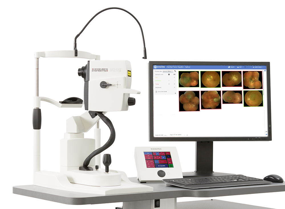
The SPECTRALIS® is an ophthalmic imaging platform with an upgradable, modular design. This platform allows clinicians to configure each SPECTRALIS to the specific diagnostic workflow in the practice or clinic.
Multimodal imaging options include: OCT, multiple scanning laser fundus imaging modalities, widefield and ultra-widefield, scanning laser angiography and OCT angiography.
These images can then be used in HSA software to highlight tumor cells accurately and in a short-time.
OptosAdvance
OptosAdvance is a comprehensive image management solution for eyecare. It enables clinicians to review, annotate, securely refer and archive images from many eyecare diagnostic devices in their practices using a single, industry-standard DICOM solution.
This along with the AI technology incorporated within the HSA software will allow health care professionals to use both software swiftly and get accurate results efficiently.
Note: This website will be updated in future.